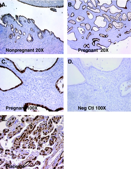
Figure 5.
Immunohistochemical localization of 17βHSD type 2 in the human cervixand placenta. Positive (brown) staining for 17βHSD type 2 is seen in the epithelial cells of the nonpregnant cervix (A) and cervix before labor (B and C). No staining is observed when the cervix is incubated without primary antibody (D). Positive 17βHSD type 2 staining is seen in the endothelial cells of the placenta (E). Magnification of A and B, 20×; C–E, 100×. Neg Ctl, Negative control.

