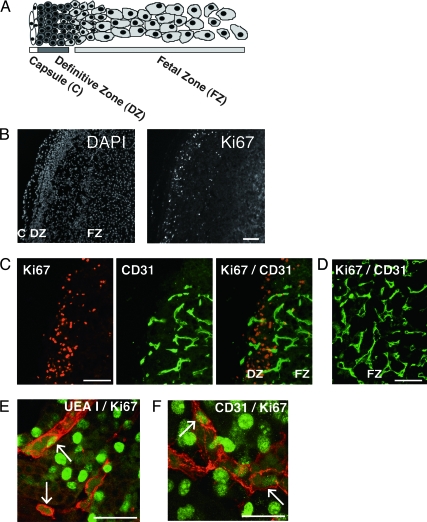
Figure 2.
Proliferating endothelial cells in the HFA gland. A, Schematic illustration of the morphology of the HFA. B, Low-power views spanning the DZ and FZ of a 15-wk HFA are shown. Nuclear staining with 4, 6-diamidino-2-phenylindole (DAPI) or immunostaining with antibody against the proliferation marker, Ki67. Ki67-immunoreactive nuclei were almost exclusively restricted to cells at the periphery of the gland. C and D, Immunostaining of a 18-wk HFA gland with anti-Ki67 (red) and anti-CD31 (green) antibodies. Low-power views spanning the periphery of the gland (i.e. the outer DZ and DZ/FZ border; C) and the inner FZ (D). E and F, Confocal microscopy images illustrating labeling for Ki67, and UEA I (UEA I) or CD31 in the DZ. Green indicates Ki67-positive nuclei. Red indicates UEA I-positive (E) or CD31-positive cells (F). Note costaining for Ki67 in the nuclei, and UEA I or CD31 on the cell membrane, indicating proliferating endothelial cells (arrows). Images are from a 17-wk adrenal (E and F). Magnification bar, 100 μm (B, C, and D) and 50 μm (E and F). Original magnification, ×100 (B), ×200 (C and D), and ×630 (E and F).

