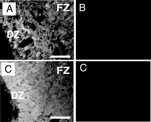
Figure 3.
VEGF-A and FGF-2 protein expression in the HFA gland. A–D, Immunofluorescence of 17-wk (A and B) and 18-wk (C and D) gestation HFAs showing localization of VEGF-A and FGF-2, respectively. VEGF-A protein (A) localized throughout the gland. FGF-2 protein (C) localized principally in the outer region of the gland. B, Control slide incubated with normal rabbit serum. D, Control slide incubated with normal mouse IgG. Magnification bar, 100 μm. Original magnification, ×200.

