Figure 6.
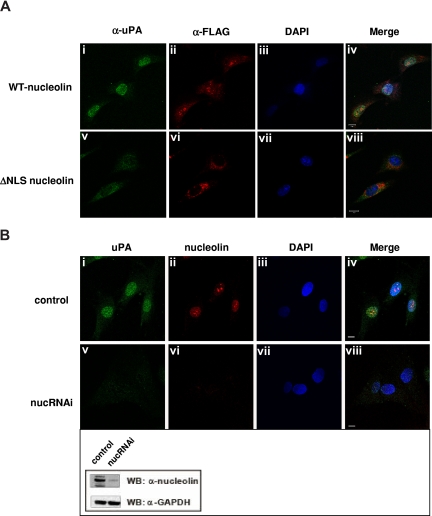
Nucleolin mediates transport of scuPA to the nucleus. (A) ΔNLS-nucleolin abrogates nuclear translocation of ΔGFD-scuPA. MEF-1 cells, transfected with vectors encoding either mouse WT-nucleolin-FLAG (top panel) or ΔNLS-nucleolin-FLAG (bottom panel), were incubated with ΔGFD-scuPA (20 nM) for 30 minutes at 37°C and then fixed in MeOH as in Figure 1. ΔGFD-scuPA was visualized using polyclonal α-uPA Ab and Alexa488-conjugated secondary Ab (green; i,v). FLAG-tagged nuceolin variants were visualized with Cy3-conjugated mouse α-FLAG MAb (red; ii,vi). Nuclei were stained with DAPI (blue; iii,vii). Panels iv and viii show merged images. (B) Effect of nucleolin down-regulation on nuclear translocation of ΔGFD-scuPA. BJ cells were transduced with “empty” lentivirus (top panel) or lentivirus delivering a cassette expressing a nucleolin-targeting shRNA (bottom panel, nucRNAi). Cells were incubated with 20 nM ΔGFD-uPA for 30 minutes, fixed, and stained as above, except that endogenous nucleolin was detected using mouse α-nucleolin MAb and Alexa546-conjugated α-mouse Ab (red, ii,vi). Panels iv and viii show merged images. Scale bar represents 10 μm. (Inset) WB of lysates from cells transduced with lentivirus variants as in panel B using mouse α-nucleolin MAb to analyze nucleolin content and α-GAPDH MAb for the control of total protein loading. A decrease in expression of nucleolin, but not GAPDH, is seen.

