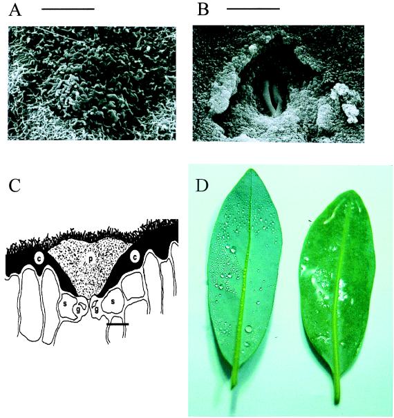
Figure 1.
Scanning electron micrograph (A) and line drawing (C) of a Drimys winteri stoma occluded by a cutin plug showing the locations of the plug, cuticle, epicuticular wax, guard cells, and subsidiary cells, and a scanning electron micrograph (B) of a leaf with the plug experimentally removed. (D) Comparison of the leaf-surface wettability of a leaf with (Left) and without (Right) stomatal plugs. p, stomatal plug; c, cuticle; g, guard cell; s, subsidiary cells. (Bars, 20 μm.)

