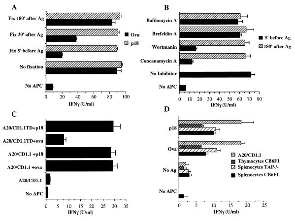
Figure 2.
Antigen processing and endosomal localization are required for Ova presentation by mCD1. (A) Fixed cells can present p18 peptide but not Ova. RMA-S/CD1.1 transfectants were fixed with 0.03% glutaraldehyde either before, or at different time points after, addition of antigens. The antigens were either 50 μg/ml Ova or 4 μg/ml p18 peptide. (B) RMA-S/CD1.1 transfectants were incubated with the indicated inhibitors: 20 nM Concanamycin A, 50 nM Bafilomycin, 200 nM Wortmannin, or 10 μg/ml Brefeldin A. Inhibitors were added either 5 min before, or 180 min after, addition of 50 μg/ml Ova. After 4 hr in the presence of the inhibitors, the APC were fixed and tested for their ability to stimulate Ova reactive T cells. (C) A20/CD1.1 and A20/CD1.1TD transfectants were pulsed either with 50 μg/ml Ova or 4 μg/ml p18 peptide for 3 hr, were irradiated, and were added to T cell stimulation assays. ELISAs were used to measure the IFN-γ levels after 3 days. In the case of A and B, the in vitro activated CTLs were derived from C57BL/6 mice whereas in C the CTLs were derived from CB6F1 mice. The control production of cytokine from untransfected cells pulsed with Ova, or T cells alone, in each case was <6 units/ml. These data are representative of one of six experiments. (D). Normal APC can present Ova to mCD1-restricted T cells. Ova-reactive T cells were generated from CB6F1 DNA immunized mice and were restimulated with Ova plus mCD1 RMA-S transfectants as described above. The representative data from one of six animals analyzed in this way is shown.

