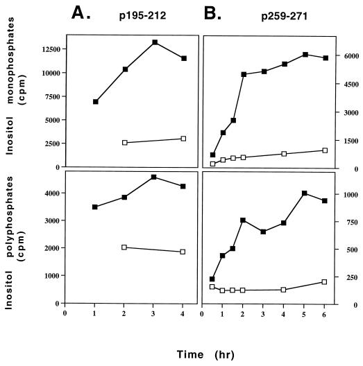
Figure 2.
Kinetics of induction of inositol phospholipid hydrolysis by the myasthenogenic peptides in the specific T cell lines. Cells of the peptide specific lines were prelabeled with myo-[2-3H]inositol and were incubated with APCs in the presence or absence of either p195–212 (15 μM) (A) or p259–271 (8 μM) (B) for the indicated time points. The levels of inositol phosphates were measured as described in Materials and Methods. The experiment is a representative of two independent experiments. □–□, without peptide; ■–■, in the presence of peptide.

