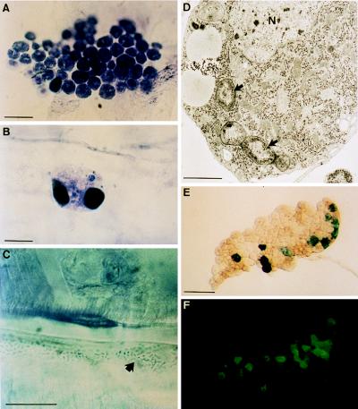
Figure 1.
Blood cells and fat body in domino larvae. Whole mounts of integument inner wall from wild-type (A) and domino (B and C) third-instar larvae. Groups of sessile plasmatocytes are frequently observed in wild-type larvae (A). However, in domino, a few abnormal hemocytes are occasionally seen (B), and microorganisms (arrow) accumulate in the hemocoele (C). Histochemical analysis showed the presence of toluidine and eosin. (For A–C, bars = 20 μm.) (D) Transmission electron micrograph of a plasmatocyte that has engulfed several bacteria (arrows). The experimental procedure was performed as described (21). N identifies the nucleus. (Bar = 1 μm.) Expression of antimicrobial reporter genes (E, diptericin-lacZ and F, drosomycin-green fluorescent protein) in a domino fat body lobe (in the absence of immune challenge). (Bar = 400 μm.)

