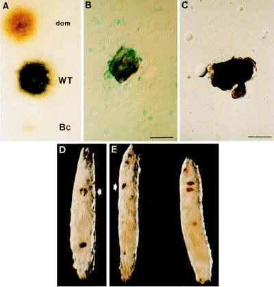
Figure 3.
Melanization and melanotic tumors in domino larvae. (A) Staining obtained 5 min after depositing a droplet of hemolymph from domino, OregonR (WT), and Bc larvae on a filter paper impregnated with l-3,4-dihydroxyphenylalanine, the substrate for phenoloxidase. Free-floating melanotic masses in Toll10B/+ single (B) and dom/dom; Toll10B/+ double mutants (C). In single mutants, the melanotic tumors are surrounded by layers of lamellocytes, as shown by the expression of the lacZ reporter of the l(3)03349 fly line (21), whereas no blood cells are associated in double mutants. (Bars = 40 μm.) (D) hopTum-l; dom/dom double-mutant larva exhibiting both the domino phenotype with black lymph glands (arrow) and a melanotic mass in the posterior region. (E) Natural infection of a domino larva by B. bassiana spores. On the left, the arrow indicates the lymph glands, and the larva on the right is an OregonR larva. Brown spots form at the penetration points of the fungi.

