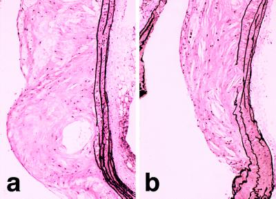
Figure 2.
Photomicrographs of atherosclerotic lesion in the aortic arch of apoE-deficient mice. Advanced intimal lesion at the root of the carotid artery in an untreated apoE-deficient mouse exhibiting necrosis, calcification, and cholesterol crystals (a). Lesion at the same location in an apoE-deficient mouse after 30 wk of ETA receptor blockade (b). Cellular composition and morphology of the plaques was similar to that seen in untreated animals. (Elastica van Giesson stain, original magnification X60).

