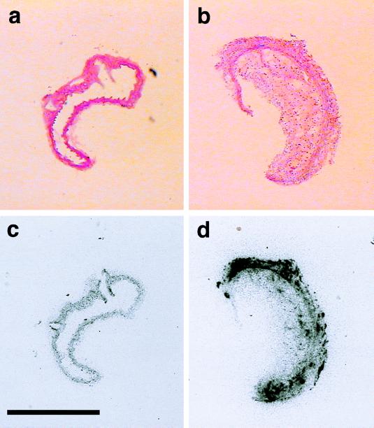
Figure 3.
Photomicrographs and autoradiographs of the aorta from C57BL/6 (Left) and apoE-deficient mice (Right). Representative histologic sections from the aorta of a C57BL/6 control mouse (a) and an atherosclerotic apoE-deficient mouse (b). Note the large plaque occluding the lumen in the aorta of the apoE-deficient mouse (b). In these animals, endothelin binding by using [125I]ET-1 was markedly increased as compared with C57BL/6 mice (c) and was localized both in the vascular wall and in the atheromatous plaque (d). (Hematoxilin/eosin stain, original magnification X10. Bar: 1,000 μm).

