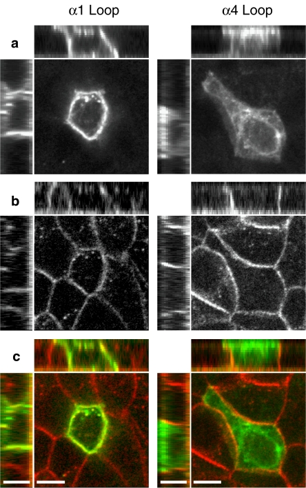
Figure 5.
Targeting of loop chimaeras examined in polarized epithelial (MDCK) cells. Representative confocal images are shown illustrating a non-polarized distribution of the α1 loop chimaera (left panels) and a polarized (apical) distribution of the α4 loop chimaera (right panels). The main images are single confocal section from 15–17 separate 0.5 μm sections. Above and to the left of the main images are representative X–Z and Y–Z confocal sections from each image in which the apical cell surface is located at the top and to the left, respectively. (a) Labelling of subunit chimaeras with Alexa-488 αBTX. (b) Antibody (rhodamine) staining of endogenous E-cadherin, which is located on the basolateral membrane of MDCK cells. (c) Merged images from (a and b) in which Alexa-488 αBTX staining is shown in green and antibody staining of endogenous E-cadherin is shown in red. Scale bars=7 μm. αBTX, α-bungarotoxin; MDCK, Madin–Darby canine kidney.

