Figure 1.
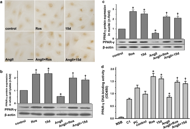
Expression and activation of PPAR-γ in cardiac fibroblasts. Cells were pretreated with or without rosiglitazone (Ros; 5 μM) or 15d-PGJ2 (15d; 5 μM) for 1 h and subsequently stimulated with Ang II (0.1 μM) for 24 h. (a) A representative immunocytochemical staining of PPAR-γ in cardiac fibroblasts (n=5; bar=50 μm). (b) Detection of PPAR-γ protein in whole-cell lysates by western blot analysis. (c) Detection of PPAR-γ protein in nuclear extracts by western blot analysis. (d) PPAR-γ activation was analysed by DNA-binding assay using PPAR-γ transcription assay kit. NSB indicates nonspecific binding; C1 for competitor; PC for positive control. All the data shown here are the mean±s.e.mean of three independent experiments. *P<0.05 vs control; †P<0.05 vs Ang II. Ang II, angiotensin II, 15d-PGJ2, 15-deoxy-Δ12,14-prostaglandin J2; PPAR-γ, peroxisome proliferator-activated receptor-γ.

