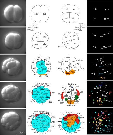
Fig. 2.
First cleavages in the ontogeny of the tunicate Oikopleura dioica. (Left) Projections of the upper (animal) half of DIC-stacks recorded for 4D microscopy. (Line drawings in Left) View from animal pole. (Line drawings in Right) View from vegetal pole. Asterisk marks the position of the blastopore. (Right) 3D representation of the positions of the nuclei of each cell in the 4D microscopy software SIMI°BioCell as seen from the vegetal pole. Cubes derive from the left side (AB2), spheres from the right (AB2). Nomenclature and color code for tissues that are already restricted as in Fig. 1. Dashed line indicates axis of bilateral symmetry. Note bilateral symmetry in these early embryonic stages.

