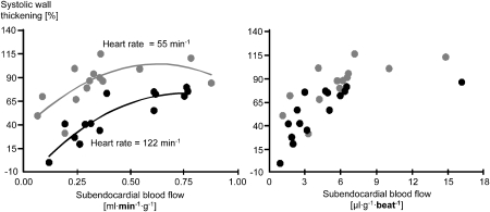
Figure 7.
Left: when heart rate is reduced by a pharmacological agent, the relationship between systolic wall thickening and myocardial blood flow is displaced leftwards and upwards, indicating that contractile function for each level of blood flow is increased. Right: when myocardial blood flow is calculated for each single cardiac cycle, the relationships at two different heart rates are superimposable, again supporting the concept of perfusion–contraction matching (Indolfi et al., 1989).

