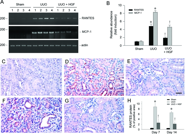
Figure 3.
HGF inhibits renal RANTES and MCP-1 expression in the obstructed kidney after UUO. A and B: RT-PCR demonstrated renal RANTES and MCP-1 mRNA expression in different groups as indicated. Numbers (1 to 4) in A denote each individual animal. Relative RANTES and MCP-1 mRNA abundance (fold induction over sham controls) was calculated after normalizing with β-actin. B: Data were presented as mean ± SEM of four animals per group (n = 4). *P < 0.05 versus sham control. †P < 0.05 versus pcDNA3. C–G: Immunohistochemical staining demonstrated that RANTES protein was induced predominantly in renal tubular epithelium in the obstructed kidney at 7 (D) and 14 (F) days after UUO, compared with sham control (C). Tubular expression of RANTES was primarily suppressed by HGF at 7 (E) and 14 (G) days after UUO, respectively. H: Graphical presentation shows the quantitative determination of RANTES protein expression in different groups as indicated. Data were presented as mean ± SEM of five animals per group (n = 5). *P < 0.05 versus sham control. †P < 0.05 versus pcDNA3. Scale bar = 20 μm.

