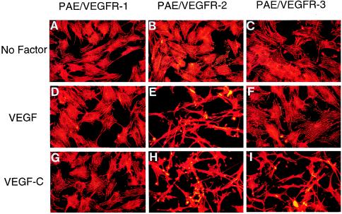
Figure 5.
VEGF-C- and VEGF-induced endothelial cell shape changes and actin reorganization. VEGF-C and VEGF were added to approximately 70% confluent VEGF receptor-expressing PAE cells at a final concentration of 100 ng/ml for 12 h, fixed with paraformaldehyde, permeabilized with Triton X-100, and stained with rhodamine-phalloidin. In response to VEGF-C (G–I) and VEGF (D–F), PAE/VEGFR-2 cells (H and E) undergo dramatic spindle-like shape changes, leading lamellae, lamellipodium extensions and actin reorganization. PAE/VEGFR-3 cells also show this striking cell shape change in response to VEGF-C stimulation (I). PAE/VEGFR-1 cells lack such a cell shape change in the presence of VEGF-C (G) and VEGF (D). All VEGFR-expressing cells also lack cell morphological changes in the absence of growth factors (A–C).

