Figure 3.
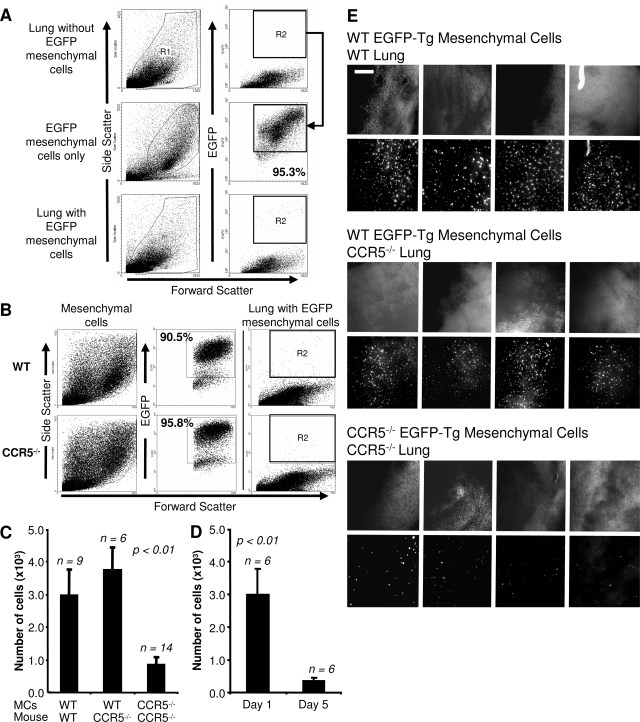
Wild-type mesenchymal cells migrate more efficiently in vivo than CCR5−/− cells. A: Gating strategy for the detection of injected EGFP-Tg mesenchymal cells. R1 includes viable cells based on FSC/SSC. R2 excludes all EGFP-Tg-negative cells based on flow cytometry analysis of an uninjected lung. These gates detect more than 95% of all EGFP-Tg cells before injection (middle). Migrating mesenchymal cells are detected in R2 after injection (bottom). B: Expression of EGFP-Tg is equivalent in wild-type and CCR5−/− mesenchymal cells. Representative FSC/SSC and FSC/EGFP-Tg dot plots of wild-type and CCR5−/− EGFP-Tg mesenchymal cells. C: More wild-type than CCR5−/− cells are detected 24 hours after intravenous injection. Bar graphs show the number of EGFP-Tg cells recovered from the right lung as determined by the percentage of EGFP-Tg cells times the number of cells isolated. The number of animals for each experiment is given above the graph. D: The number of detected wild-type fibroblasts decreases throughout time. A similar strategy as described in C was applied to mice 1 and 5 days after injection. E: Fluorescent microscopic images of the left lungs of mice from C using a SteREO Lumar V dissecting scope. Scale bar = 1 mm. Original magnifications, ×42.

