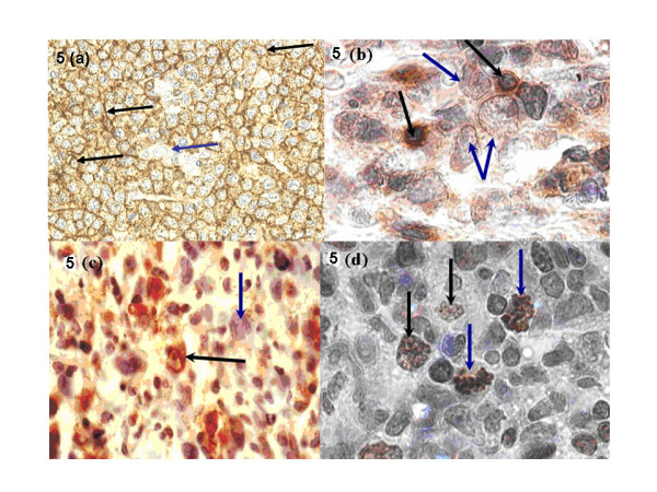
Figure 5.

a) Anti-CD20 (B-cell) antigen immunoperoxidase reactivity in DLBCL; note the granular cytoplasmic reactivity (brown) in all malignant cells [black arrows] while a macrophage did not stain [blue arrow] (× 200). (b) Anti-CD3 (T-cell) antigen immunoperoxidase reactivity in DLBCL; note the brown focal cytoplasmic reactivity in tumor infiltrating T-lymphocytes [black arrows] while the malignant cells were not reactive [blue arrows] (× 400). (c) Anti-CD68 antigen immunoperoxidase reactivity in DLBCL; note the brown cytoplasmic reactivity in tumor infiltrating macrophages [black arrow] while malignant cells did not stain [blue arrow] (× 400). (d) Anti-Ki-67 (proliferation) antigen immunoperoxidase reactivity in an aggressive lymphoma: note the brown granular nuclear reactivity in proliferating cells [black arrows] including the abnormal mitotic bodies [blue arrows] (× 400).
