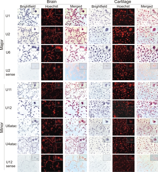
Fig. 1.
Localization of spliceosomal snRNAs in embryonic mouse tissues. ISH was performed on sagittal sections of E15 mouse embryos using DIG-labeled full-length RNA probes complementary to the snRNAs indicated. NBT/BCIP staining was performed for 40 min (major snRNAs) or 4 h (minor snRNAs). Blue color in the brightfield images represents the signal from the snRNA probes. Hoechst staining of nuclei in the darkfield images was converted to red by image processing. In the merged images, colocalization of the snRNA and Hoechst signals is indicated by purple. (Inset) An enlarged Inset of individual cells is included at the top right corner of each image. (Scale bar is 20 μm.)

