Figure 2.
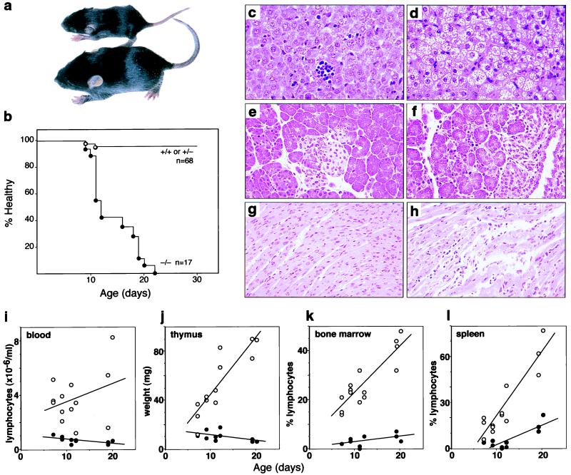
Phenotypic abnormalities in SOCS-1−/− mice. (a) SOCS-1−/− and littermate mice at 10 days of age showing retarded growth of the SOCS-1-deficient mouse. (b) Rapid onset of morbidity and mortality in SOCS-1−/− mice. The proportion of mice remaining disease-free (percent healthy) with age is shown. All of the SOCS-1−/− mice died within 3 weeks of birth. Normal histological appearance of the liver (c), pancreas (e), and heart (g) of littermate mice is in contrast to the fatty degeneration of the liver (d) and monocytic infiltration of the pancreas (f) and heart (h) of SOCS-1−/− mice. The progressive deficit with increasing age of SOCS-1−/− mice (•) in peripheral blood lymphocytes (i), thymic weight (j), and lymphocyte composition of the bone marrow (k) and spleen (l) is shown in comparison with values from littermate SOCS-1+/+ or SOCS-1+/− controls (○).

