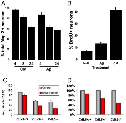
Fig. 3.
The behavior of E16.5 neurons (5 DIV) after Ab1–42 treatment. (A) Both CM and Ab1–42 (Aβ) lead to loss of Map2+ neurons from the culture. Counts are shown at 4, 8, and 24 h after Aβ addition. (B) Proportion of BrdU/Map2 double-positive cells after the indicated treatment. The absence of Cdk5 does not protect from Aβ-induced cell death. (C) Total cells per field in both Aβ-treated (red bars) and untreated (gray bars) cultures. The absence of Cdk5 leads toTuJ1+ cell loss after 24 h (6), but the effect of Aβ can be seen on top of this. (D) When expressed as a percentage of the control cultures, the percentage loss is higher in Cdk5−/− cultures, arguing against a pathogenic role for Cdk5 in neurodegeneration.

