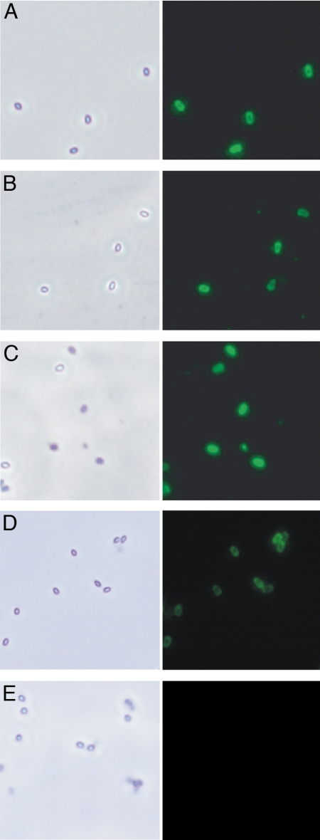
Fig. 2.
Immunofluorescent staining of B. anthracis spores. (Left) Phase-contrast microscopy showing the spores. (Right) Spores treated with hyperimmune anti-Shewanella spp. MR-4 (A); anti-Conjugate no. 1: BSA/CPSTSB (B); anti-Conjugate no. 2: BSA/CPSCDM (C); hyperimmune anti-P. syringae (D); and preimmune control serum (E).

