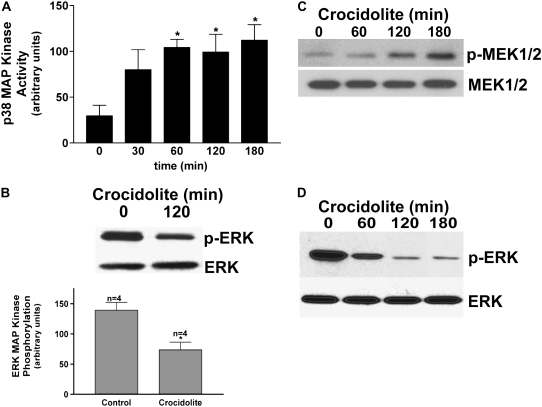
Figure 2.
Differential MAP kinase activation in human monocytes exposed to crocidolite asbestos. (A) Whole cell lysates were prepared from human monocytes (n = 3) exposed to crocidolite asbestos for the indicated amount of time. The lysates were subjected to immunoprecipitation with the p38 MAP kinase polyclonal antibody. In vitro kinase assays were performed using ATF-2 as the substrate. Western blot analysis for p38 MAP kinase was performed to confirm equal loading of the proteins. *P < 0.019. (B) Human blood monocytes (n = 4) were cultured with or without crocidolite asbestos for 2 hours. Western blot analysis was performed for p-ERK monoclonal and ERK polyclonal antibodies to determine activation and confirm equal loading of the proteins, respectively. Densitometry was performed from the four experiments and is expressed graphically in arbitrary units. For statistical comparisons, * denotes a comparison of p-ERK in blood monocytes cultured with and without crocidolite asbestos (P < 0.0057). (C) Human blood monocytes were exposed to crocidolite asbestos for the indicated amount of time. Western blot analysis was performed with p-MEK1/2 monoclonal and MEK1/2 polyclonal antibodies to determine activation and confirm equal loading of the proteins, respectively. (D) Human blood monocytes were exposed to crocidolite asbestos for the indicated amount of time, and whole cell lysates were incubated with an active ERK kinase for 20 minutes at 37°C, as described in Materials and Methods. Samples were separated by SDS-PAGE and Western analysis was performed with p-ERK monoclonal and ERK polyclonal antibodies.

