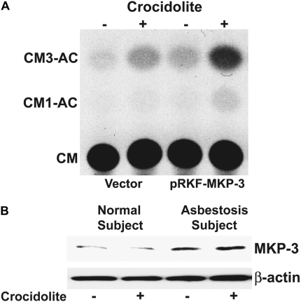
Figure 6.
MKP-3 augments TNF-α gene expression in monocytes exposed to asbestos. (A) THP-1 cells were transiently transfected with the −600 TNF-α-CAT reporter plasmid and either an empty vector or pRKF-Flag-MKP-3 expression vector. After 24 hours, cells were exposed to crocidolite asbestos for 24 hours. Whole cell lysates, which were normalized to protein, were incubated with 0.1 μCi of [14C] chloramphenicol and 1.0 mM acetyl coenzyme A for 2 hours at 37°C. Acetylated derivatives (CM3-AC and CM1-AC) were separated from nonacetylated chloramphenicol (CM) by ascending thin layer chromatography in chloroform/methanol (95:5) solvent. (B) Whole cell lysates were prepared from alveolar macrophages obtained from normal subjects (n = 2) or from patients with asbestosis (n = 2) cultured for 1 hour in the presence or absence of crocidolite asbestos. Lysates were separated by SDS-PAGE. Western blot analysis was performed using the MKP-3 polyclonal and β-actin monoclonal antibodies to determine expression and equal loading of the proteins, respectively. A representative Western blot analysis is shown.

