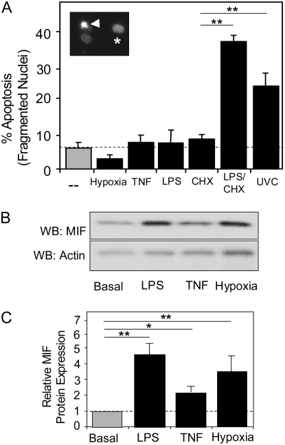
Figure 1.
The response of human pulmonary artery endothelial cells (HPAEC) to potential apoptotic stimuli in culture. (A) Multiple cytotoxic stressors were evaluated for their ability to induce apoptosis in HPAEC. Basal apoptosis (dotted line) is minimal, representing less than 5% of the total cells evaluated under the culture conditions detailed in Materials and Methods. To induce hypoxia, a mixture of nitrogen and carbon dioxide (95%/5%) gases were used to purge cultures of oxygen for 48 hours before quantification of apoptotic cells. Additional cultures were incubated in the presence of TNF (100 ng/ml), lipopolysaccharide (LPS; 100 ng/ml), cycloheximide (CHX; 50 μg/ml), or LPS/CHX (100 ng/ml and 50 μg/ml, respectively) for 24 hours before analysis. Insert shows Hoechst 3342 staining of normal (asterisk) and fragmented (arrowhead) nuclei. (B) Macrophage migration inhibitory factor (MIF) protein and β-actin were identified by Western blotting in total cell lysates of untreated HPAEC and LPS-, TNF-α–, and hypoxia-treated HPAEC. (C) Graphical representation of intracellular MIF protein levels normalized to β-actin expression. Results from at least three independent experiments. (*P > 0.01; **P > 0.005).

