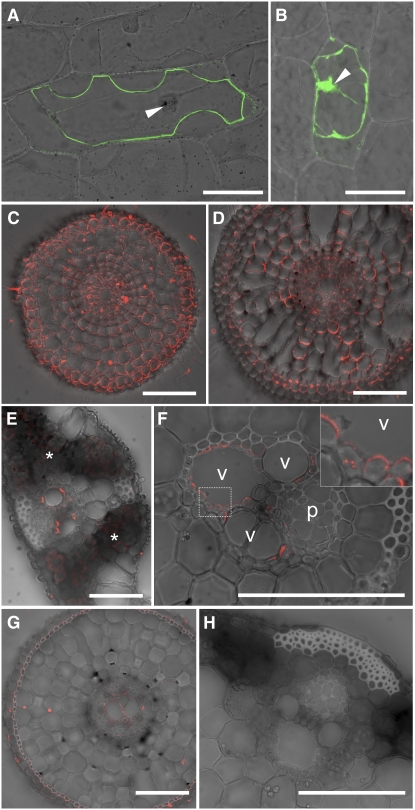
Figure 3.
Subcellular and Tissue-Specific Localization of Lsi6 Protein.
(A) and (B) Transient expression of a GFP-Lsi6 fusion (A) and GFP alone as control (B) introduced by particle bombardment into onion epidermal cells. The fluorescence was observed after plasmolysis of cells with 1 M mannitol. Merged images of confocal GFP fluorescence and bright field are shown. Arrowheads indicate the nuclei.
(C) to (F) Immunostaining of Lsi6 protein in wild-type rice. Seminal root cross sections 5 and 30 mm from the tip ([C] and [D], respectively), leaf blade (E), and leaf sheath (F) with an enlarged image of xylem parenchyma (inset, corresponding to the frame in [F]) are shown. Fluorescence from secondary antibodies labeled with Alexa Fluor 555 is shown in red. (C) and (D) were stained and observed under identical conditions on the same slide. Asterisks in (E) indicate weak autofluorescence from chloroplasts in mesophyll cells. Vessel and phloem are indicated as v and p, respectively, in (F).
(G) and (H) Anti-Lsi6 antibody immunostaining in the Lsi6 T-DNA insertion line. A root cross section at 30 mm from the tip (G) and a leaf sheath (H) are shown.
Bars = 100 μm.

