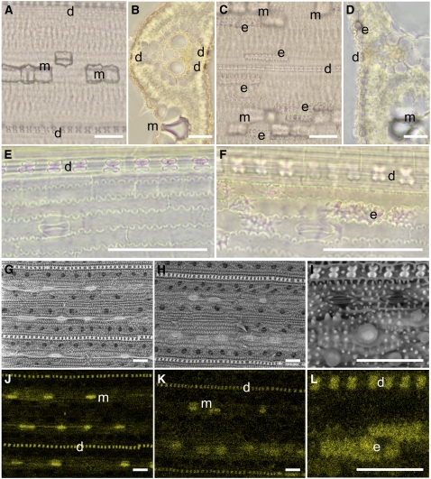
Figure 6.
Silica Distribution in the Leaf Blade.
(A) to (F) Phenol-safranin staining of leaf blade ([A] to [D]) and leaf sheath ([E] and [F]) of wild-type cv Dongjin ([A], [B], and [E]) and the Lsi6 T-DNA insertion mutant ([C], [D], and [F]) after 0.5 mM Si treatment for 6 d. Longitudinal ([A], [C], [E], and [F]) and cross sections ([B] and [D]) were observed by optical microscopy.
(G) to (L) Images from scanning electron microscopy coupled with EDX spectroscopy. Leaf blade of cv Dongjin ([G] and [J]) and T-DNA line ([H], [I], [K], and [L]) treated with 0.5 mM Si for 6 d were observed by scanning electron microscopy ([G] to [I]) and simultaneously detected with EDX of Si ([J] to [L]). Yellow color in (J) to (L) indicates the characteristic x-rays from Si.
d, silicified dumbbell-shaped cell; m, silicified motor cell; e, silicified abaxial epidermis. Bars = 50 μm.

