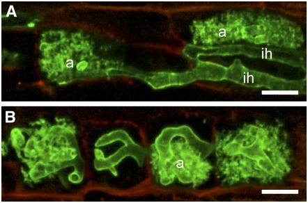
Figure 2.
Different Patterns of G. gigantea Colonization in the Inner Cortex of M. truncatula and D. carota Roots.
The AM fungal cell wall was stained using tetramethylrhodamine-conjugated wheat germ agglutinin and false colored in green to maximize the contrast with the weak red autofluorescence of the plant cell walls. Bars = 20 μm.
(A) In M. truncatula roots, terminal arbuscules (a) were formed within inner cortical cells following infection by intercellular hyphae (ih).
(B) In D. carota, intercalary arbuscules (a) were formed within adjacent intracellularly infected inner cortical cell files, and intercellular hyphae were absent. Both images are z axis projections of serial optical sections acquired from vibratome longitudinal root sections.

