Figure 5.
Nuclear Enlargement and Remodeling in Inner Cortical Cells Associated with Prepenetration Responses.
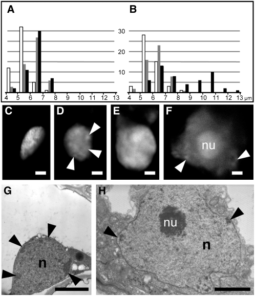
(A) and (B) Histograms showing the distribution of nuclear sizes in epidermal (white), outer cortical (gray), and inner cortical cells (black) in M. truncatula control (A) and colonized roots (B). The plots represent 50 independent measurements for each cell type. The population of significantly enlarged nuclei in colonized roots corresponded to inner cortical cells with prepenetration responses.
(C) to (F) Propidium iodide fluorescent staining of DNA to visualize changes in nuclear morphology. Control inner cortical cells of D. carota (C) and M. truncatula (E) had densely stained nuclei without visible nucleoli. By contrast, enlarged nuclei in D. carota (D) and M. truncatula (F) cells with prepenetration responses contained only isolated patches of condensed chromatin (arrowheads) at the nuclear periphery, and the nucleolus (nu) was often very large (F).
(G) and (H) TEM images of the nucleus (n) in a control (G) and AM-responding cell (H) of D. carota at the same magnification. In addition to the obvious size difference, the prepenetration-responsive nucleus (H) had lower chromatin density, fewer dark peripheral patches of heterochromatin (arrowheads), and a very large and granular nucleolus (nu). Bars = 2 μm.

