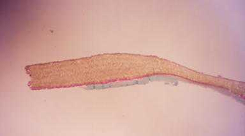
Fig. 1.
Histologic image showing the inner, nonporous surface (up) and the outer, porous surface (bottom) of a PSM that had been implanted for 6 months. While there is no scar tissue visible on the inner surface, abundant fibroblasts are present within the material on the outer surface (magnification 33× Milligan’s trichrome stain)

