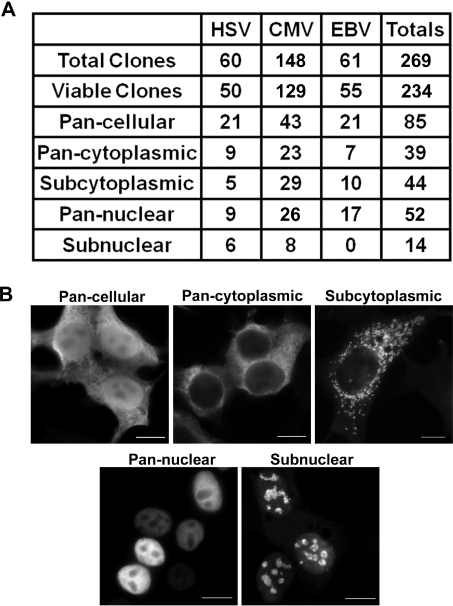
Figure 1. Localization summary of herpesvirus proteins expressed in 293T cells.
(A) Summary table indicating the localizations of herpesvirus proteins expressed in transfected 293T cells as determined by IF detection of the C-terminal FLAG epitope. Viable clones refers to the number of clones for which the viral protein was detected by IF. (B) IF micrographs representative of the pan-cellular, pan-cytoplasmic, subcytoplasmic, pan-nuclear and subnuclear localizations. Scale bar = 10 µm.

