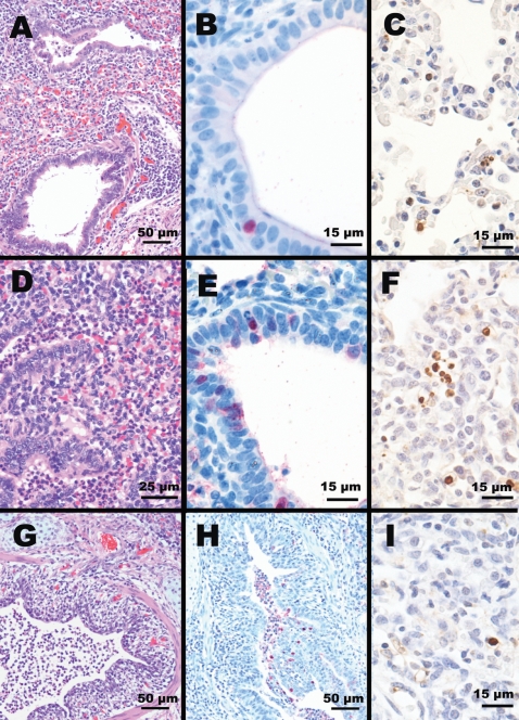
Figure 5. Histopathologic findings in lungs of pigs on day 5 after virus inoculation.
(A) Moderate lymphocytic bronchiolitis with slight intra-luminal cellular debris (HE staining) and (B) viral antigen reaction in a single bronchiolar cell (IHC staining) in the lungs of pigs infected with VN/04 (H5N1) virus. (D) Moderate bronchioalveolitis with moderately cellular debris in the airway of bronchioles and alveoli (HE staining) and (E) marked viral antigen reaction in bronchiolar cells (IHC staining) in the lungs of pigs infected with WS/Mong/05 (H5N1) virus. (G) Severe bronchitis characterized by degeneration and necrosis of bronchi epitheliums with severe intra-luminal cellular debris (HE staining) and (H) prominent viral antigen reaction in the bronchial epitheliums and necrotic cellular debris (IHC staining) in the lungs of pigs infected with Sw/NC/88 (H3N2) virus. Apoptosis frequently observed in proliferating leukocytes and macrophages in the lungs of pigs infected with (C) VN/04 and (F) WS/Mong/05 H5N1 viruses (TUNEL assay). (I) TUNEL assay of lungs from pig infected with Sw/NC/88 (H3N2) virus–a single cell affected with apoptosis.

