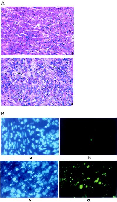
Figure 4.
(A) Established MDA-MB-468 tumors were injected with H5.110lacZ (Upper) or H5.110hIFNβ (Lower) at 1 × 10⁹ pfu per mouse, and tumors were harvested 4 days later. Histological analysis then was performed; hematoxylin/eosin-stained sections are shown. More apoptotic cells (indicated by the arrows) were noted in the H5.110hIFNβ-injected tumor. (B) Confirmation of apoptosis by 4′,6-diamidino-2-phenylindole staining (a and c) or direct fluorescence detection of end-labeled and fragmented genomic DNA (b and d) in tumors treated either with H5.110lacZ (a and b) or H5.110hIFNβ (c and d).

