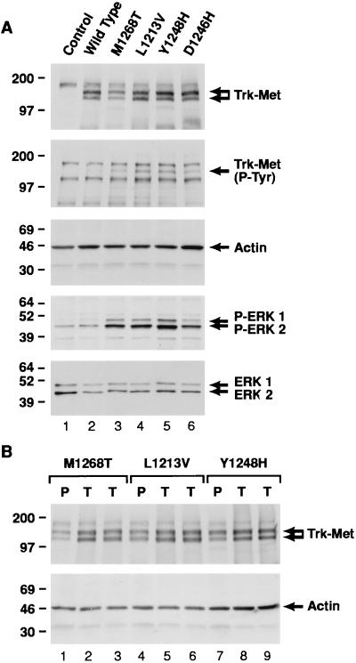
Figure 1.
Western analysis of NIH 3T3 cells expressing mutationally activated Trk-Met. (A) Samples labeled control and wild type are from cells stably transfected with empty vector or vector expressing wild-type Trk-Met, respectively. All other samples are from cells stably transfected with vectors expressing Trk-Met harboring the indicated activating Met mutation. Cells were cultured in DMEM/10% CS supplemented with 800 μg/ml G418, and 50 μg of cell lysate/sample was resolved on a 4–15% gel and examined under nonreducing conditions (top three blots). Cells were cultured in DMEM/1% CS for 24 hr, and 10 μg of cell lysate/sample was resolved on a 10% gel and examined under reducing conditions (bottom two blots). The following primary antibodies were used (top to bottom): anti-Met, antiphosphotyrosine, anti-actin, antiactive (i.e., phosphorylated) ERK1/2, and anti-ERK1/2. (B) Samples labeled with a “P” are from a pool of cells stably transfected with Trk-Met harboring the indicated activating Met mutation, which were used as the inoculum for tumor induction. Samples labeled with a “T” are explanted tumors derived from cells expressing the indicated Trk-Met mutant. Cells were cultured in DMEM/10% CS supplemented with 800 μg/ml G418, and 50 μg cell lysate/sample was resolved on a 4–15% gel and examined by Western analysis under nonreducing conditions using anti-Met (Upper) or anti-actin antibody (Lower). Molecular mass markers are indicated on the left.

