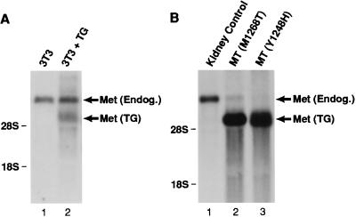
Figure 3.
Transgene expression in mammary carcinomas induced by mutationally activated Met. Fifteen micrograms of total RNA/sample was resolved on a 1.2% agarose-formaldehyde gel and examined by Northern analysis by using an anti-Met probe. (A) RNA was obtained from parental NIH 3T3 cells (lane 1) or NIH 3T3 cells stably transfected with wild-type Met under the control of the metallothionein promoter (lane 2). Note the appearance of an ≈5-kb Met-reactive transcript [designated Met transgene (TG)] only in transfected cells. The ≈7.5-kb transcript represents endogenous Met (38). (B) RNA was obtained from normal murine kidney (lane 1) or mammary tumors (MT) from founder transgenic animals harboring the indicated activating Met mutation (lanes 2 and 3). Note the strong expression of the Met transgene (TG) in tumor tissue relative to endogenous Met in these samples and to normal kidney [a tissue that expresses high levels of Met (38)]. The migration of 28S and 18S ribosomal RNA is indicated on the left.

