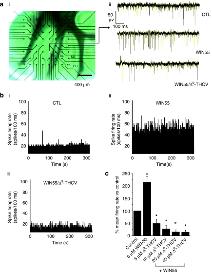
Figure 5.
Cannabinoids modulate PC spontaneous excitatory spike firing frequency in cerebellar brain slices. (a(i)) Micrograph showing a cerebellar slice adhering to an MEA. Specific cerebellar layers: GL=granular layer, ML=molecular layer and PC=Purkinje cell layer. The filled white circle shows a recording electrode position in the PC layer. (a(ii)) Examples of continuous MEA recordings from a single electrode showing effects of WIN55 (5 μM) and subsequent application of Δ9-THCV (5 μM) in the continued presence of WIN55 (5 μM) on PC control (CTL) spontaneous excitatory spike firing frequency; steady-state effects, drugs applied for a minimum of 20 min. Note the increase in PC firing rate upon application of WIN55 and consequential decrease below control levels upon subsequent application of Δ9-THCV. (b) PC spike firing rate histograms (minimum 5 min continuous recording, 100 ms bin size) for a representative electrode for (i) control, (ii) WIN55 (5 μM) and (iii) subsequent application of Δ9-THCV (5 μM). (c) Bar graph showing summarized WIN55 (5 μM) and Δ9-THCV (5–40 μM; all n=28 (6)±s.e.mean) in the continued presence of WIN55 (5 μM) effects on PC control (n=36 (6)±s.e.mean) spontaneous excitatory spike firing rates; *P<0.05 vs control; Mann–Whitney U-test.

