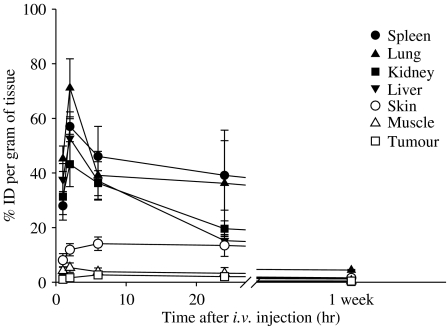
Figure 2.
Tissue distribution of BAM-SiPc. One μmol of BAM-SiPc kg−1 body weight was injected, intravenously, into the HepG2-bearing nude mice. Mice were killed at different timed intervals. Tissues were excised and homogenized with DMF. The DMF fraction was subjected to fluorescence measurement to determine the amount of BAM-SiPc, expressed as % initial dose g−1 tissue. Data are expressed as mean±s.d., n=5.

