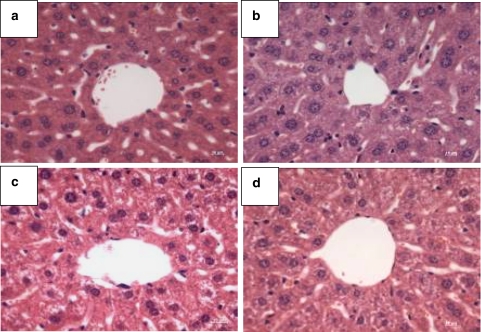
Figure 4.
Typical microscopy images obtained after H & E staining of liver sections from PDT-treated mice, showing the region around the hepatic artery. Fifteen days after PDT, livers were excised from the nude mice and sections were stained. Nuclei were stained by haematoxylin solution (purple) and the cytoplasm was counterstained by eosin solution (pink). (a) PDT-treated; (b) treated but not irradiated; (c) untreated but irradiated and (d) untreated and not irradiated. Original magnification is × 40.

