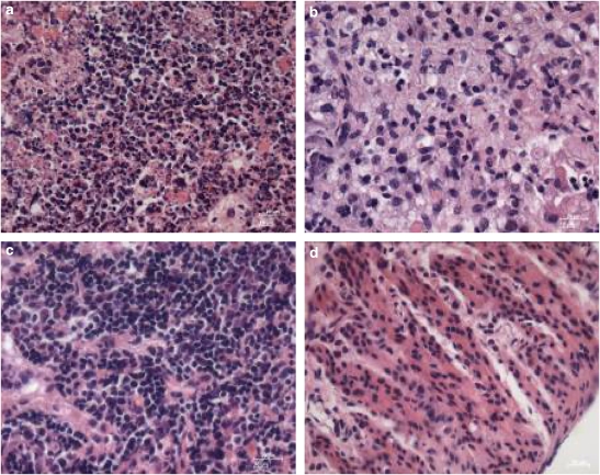
Figure 7.
Typical microscopy images obtained after H & E staining of solid tumour from PDT-treated mice. At 15 days after PDT, the HepG2 solid tumours were excised from the nude mice and sections were stained. (a) PDT-treated; (b) treated but not irradiated; (c) untreated but irradiated and (d) untreated and not irradiated. Original magnification is × 40.

