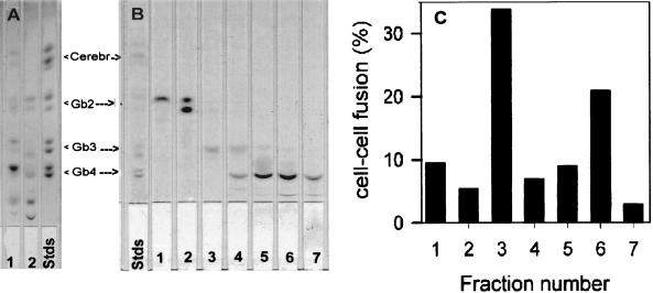
Figure 1.
Recovery of fusion after addition of various GSL fractions isolated from the crude human erythrocyte mixture. (A) TLC analysis of erythrocyte GSLs. GSLs were isolated as described in Materials and Methods. Total lipid (25–50 μg) was spotted on a 5 × 20-cm silica-gel TLC plate. The plate was developed in CHCl3/MeOH/H2O (65:25:4, vol/vol). At the end of the run, the plate was air dried, sprayed with Bial’s reagent, heated to develop the spots, scanned with a Hewlett–Packard 4P scanner, and photographed. GSLs isolated from human erythrocytes are in lane 1, and GSLs isolated from bovine erythrocytes are in lane 2; GSL standards are in lane 3. (B) TLC of various GSL fractions purified by silica-gel column. The crude GSL mixture from human erythrocytes (shown in A, lane 1) was fractionated by silica-gel column chromatography. GSLs in the fractions were analyzed by chromatography on silica-gel TLC as in A. Lanes 1–7 are fraction numbers. (C) Recovery of fusion of CD4+ nonhuman cells by GSL fractions. GP4F cells were plated on microwells and infected with vCB3 to express human CD4 on the cell surface. Liposomes containing different GSL fractions were incorporated into the membranes of the target cells and the cells were labeled with CMTMR. DiO-labeled TF228 cells were cocultured with GSL-supplemented target cells for 4–6 h at 37°C. Images were collected, and fusion was calculated as described in Materials and Methods. Data presented here are normalized to the recovery of fusion by the crude human GSL fraction.

