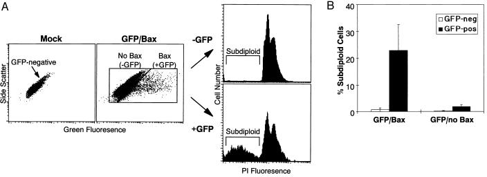
Figure 5.
Effect of transient overexpression of p21BAX on apoptosis in U343 cells. (A) U343 cells were either mock-transfected or cotransfected with two plasmid DNAs that expressed GFP and p21BAX stained with PI for DNA content 2 days posttransfection. Dot plots of green fluorescence vs. side scatter are shown. The plot describing mock-transfected cells indicates the distribution pattern of GFP-negative cells. The GFP/p21BAX cotransfected cells were separated into p21BAX-negative (GFP-negative) and p21BAX-positive (GFP-positive) populations as indicated and separately analyzed on a PI histogram. The subdiploid cell populations lie to the left of the G1 peak and are noted in each histogram. (B) Bar graph summarizing the subdiploid fraction of U343 cells 2 days after transient cotransfection with plasmids encoding GFP and p21BAX or after transient transfection with plasmid encoding GFP alone. The GFP-negative and GFP-positive populations were analyzed separately on PI histograms as detailed in A to determine the percentage of subdiploid, apoptotic cells. Each bar represents the mean ± SD of the results of three independent experiments.

