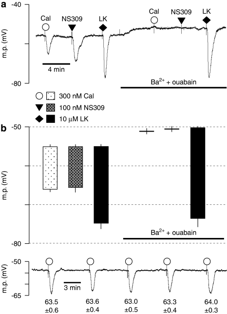
Figure 2.
Effect of 30 μM Ba2++500 nM ouabain on smooth muscle hyperpolarizations to calindol (Cal) and NS309 in Zucker lean (ZL) rat mesenteric arteries. (a) Typical trace showing abolition of the hyperpolarization to 300 nM calindol or 100 μM NS309 (6,7-dichloro-1H-indole-2,3-dione 3-oxime) by 30 μM Ba2++500 nM ouabain in a segment of artery from a ZL rat. At the end of the experiment, 10 μM levcromakalim (LK) was added to confirm the integrity of the preparation. (b) Graphical representation of mean results obtained from four experiments of the type shown in panel a. Each column represents the mean membrane potential (m.p.) before (+ s.e.mean) and after (− s.e.mean) addition of calindol, 6,7-dichloro-1H-indole-2,3-dione 3-oxime (NS309) or levcromakalim in the absence or presence of Ba2++ouabain as indicated. (c) Typical time-control experiment to test for the reproducibility of calindol-induced hyperpolarizations. After myocyte impalement, vessels were exposed to 300 nM calindol every 5 min during a 20 min impalement. Each number represents the maximum mean m.p.±s.e.mean (n=5 vessels) recorded in the presence of 300 nM calindol. Over the 20 min duration of the experiments, there was no time-dependent change in the magnitude of calindol-induced hyperpolarizations (repeated measures ANOVA; P=0.29).

