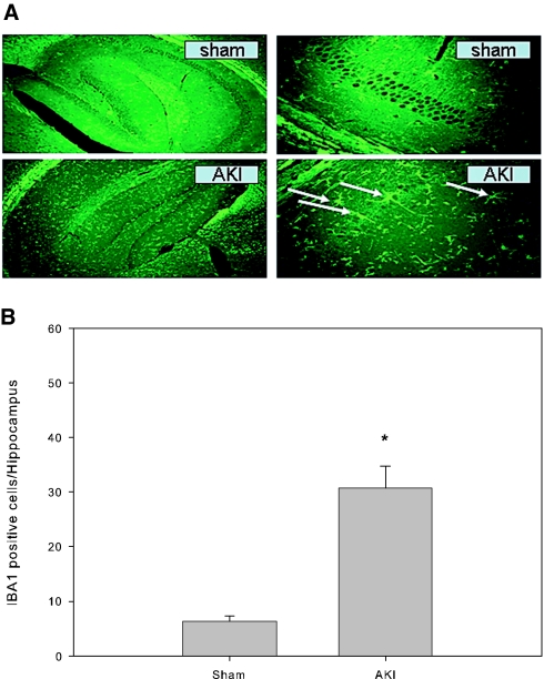
Figure 2.
Microglial cells in the hippocampus of mouse brain. All mice underwent either 60 min of renal ischemia followed by reperfusion or a sham operation. The brains were harvested at 24 h after surgery and stained with IBA1 antibody for activated microglial cells (macrophage in the brain) by immunofluorescence technique. (A) Representative microphotographs of the hippocampus. Arrows indicate positively stained microglia. (B) IBA1-positive microglial cell count in the hippocampus. *P = 0.001 versus sham; n = 6 to 7. Magnifications: ×4 in A, left; ×40 in A, right.

