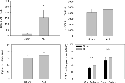
Figure 6.
Liver function, blood inflammation, and brain changes in mice with ischemic ALI. Mice underwent either 45 min of lobar ischemia followed by reperfusion or a sham operation. The blood was taken at 24 h after surgery and measured for ALT and CRP. The brains were processed for histology and GFAP staining. Mice with liver ischemia had significant increase in serum ALT meanwhile with a comparable serum CRP, brain pyknotic neuronal cell count, and brain GFAP expression when compared with sham operated mice. (*P = 0.016 versus sham; n = 4 to 5). Sham versus ALI, serum CRP: 4116.154 ± 541.709 versus 4625.344 ± 642.728 ng/ml (P = 0.213; n = 5); pyknotic neurons: 30.4 ± 3.3 versus 33.9 ± 4.5 ng/ml (P = 0.157; n = 5 to 9); GFAP-positive pixel analysis: corpus collosum 32619 ± 2715 versus 39379 ± 8890 ng/ml (P = 0.173); cerebral cortex 53833 ± 9071 versus 62383 ± 9802 ng/ml (P = 0.167; n = 5 to 9).

