Figure 2.

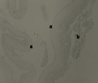
H-E × 40. Histological view of the intrahydrocelic sac revealing the internal surface lining of the mesothelial cells (a) and the attached fibrin secondary to the torsion (b).

H-E × 40. Histological view of the intrahydrocelic sac revealing the internal surface lining of the mesothelial cells (a) and the attached fibrin secondary to the torsion (b).