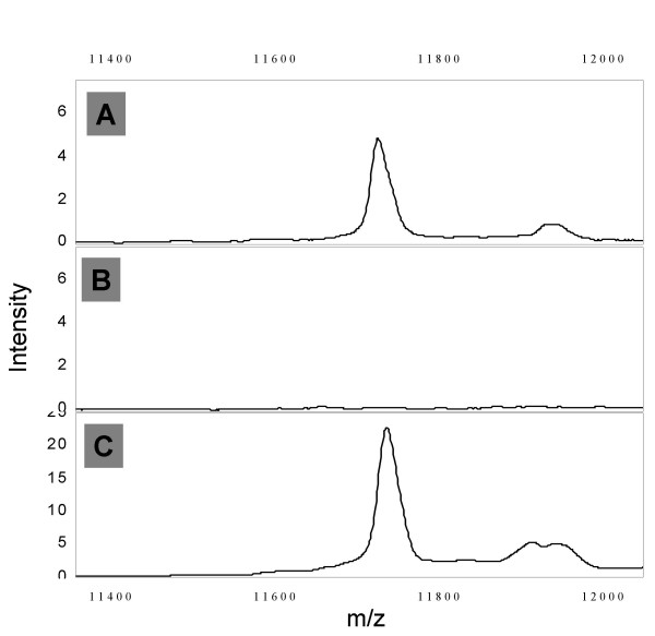
Figure 6.
β2-microglobulin immuno-MS. Panel A shows the IMAC-SELDI spectrum of neat serum, panel B the spectrum of the same serum following depletion with protein G beads loaded with rabbit polyclonal anti-serum to human β2-microglobulin (Sigma M8523) and panel C the spectrum of the eluted β2-microglobulin.

