Figure 3.
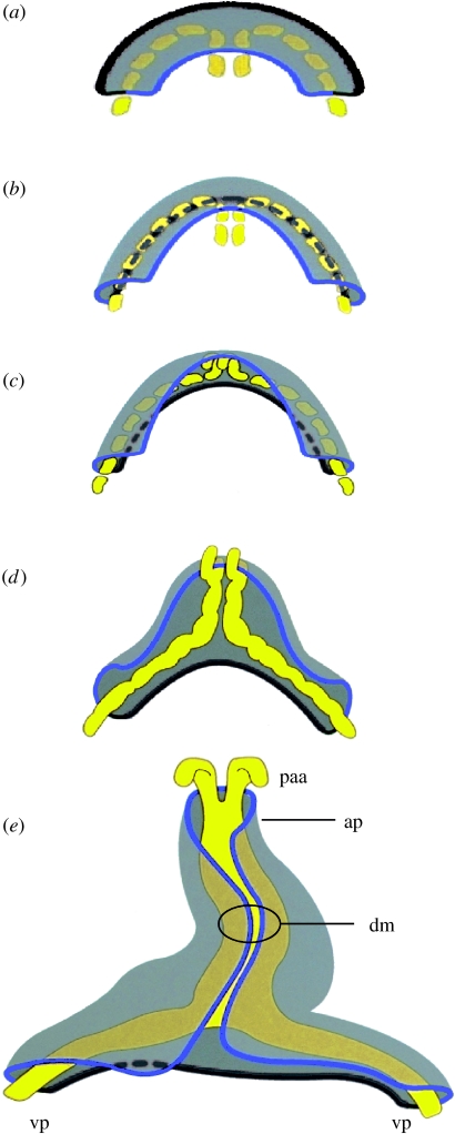
(a–e) Formation of the cardiac tube from the primary cardiac crescent, represented in five consecutive phases based on de Jong et al. (1997). See online data supplement for a link to an animation to visualize this process. The cardiac crescent is shown from the amniotic cavity (dorsal view). The other germ layers, and the rest of the mesoderm, are not shown. The myocardium is shown in transparent grey; the endocardium is shown in yellow as it forms between the endoderm and the mesodermal myocardium. The blue line marks the central border of the cardiac crescent, which is contiguous with the pharyngeal mesoderm. The dark grey line marks the peripheral border, which is contiguous with the mesoderm of the septum transversum, in which develops the systemic venous tributaries. Note that central ‘blue’ region becomes positioned dorsally, explaining the relationship of outflow components with the mediastinal component at the venous pole. paa, pharyngeal arch artery; ap, arterial pole; vp, venous pole and dm, dorsal mesocardium.

