
TABLE 1.
Data collection and refinement statistics
| Statistic | Type 1 | Type 2 | ||
|---|---|---|---|---|
| Data collection | ||||
| Space group | P1 | P1 | ||
| Unit cell dimensions (Å, degree) | a = 42.36, b = 65.84, c = 71.14 | a = 43.20, b = 63.72, c = 73.26 | ||
| α = 66.40, β = 89.93, γ = 90.04 | α = 66.64, β = 89.99, γ = 90.30 | |||
| Spacing (Å) | 20.0 − 1.80 | 1.86 − 1.80 | 29.25 − 2.00 | 2.07 − 2.00 |
| No. of measured reflections | 244,351 | 182,859 | ||
| No. of unique reflections | 62,779 | 47,224 | ||
| Average redundancy | 3.9 | 3.7 | 3.9 | 3.7 |
| Completeness (%) | 96.3 | 95.1 | 97.4 | 93.6 |
| Rmerge (I)* | 0.064 | 0.201 | 0.038 | 0.110 |
| Average I/σ (I) | 17.1 | 5.5 | 21.0 | 4.8 |
| Solvent content (%)/VM | 50.1/2.47 | 51.0/2.47 | ||
| Refinement | ||||
| No. of reflections (%) | 62,708 (96.0) | 47,200 (97.2) | ||
| No. of reflections in working set (%) | 59,547 (91.3) | 44,801 (92.4) | ||
| No. of reflections in test set (%) | 3161 (4.8) | 2399 (4.9) | ||
| R† | 0.237 | 0.249 | ||
| Free R‡ | 0.316 | 0.304 | ||
| No. of peptide chains | 4 | 4 | ||
| No. of protein/water atoms | 5118/1460 | 5096/361 | ||
| RMSD bond length (Å) | 0.008 | 0.008 | ||
| RMSD bond angle (degree) | 1.1 | 1 | ||
| Average B factor | 42.4 | 52.9 | ||
*
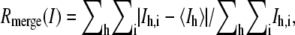 where Ih,i is the intensity of the ith observation of the reflection h.
†
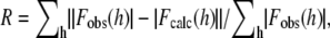 where Fobs(h) and Fobs(h) are the observed and calculated structure factors, respectively.
‡
Free R is calculated for randomly selected 5% of the reflection data, which were not included in the refinement.