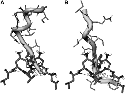
FIGURE 4.
A closeup of the dominant binding interface determined for the rigidly docked peptide NIIGRNLLTQI (shown in grayscale ribbons) in the complex with the HIV-1 PR monomer (A) and a closeup of the binding interface determined for the flexibly docked peptide (shown in grayscale ribbons)in the complex with the HIV-1 PR monomer (B). The predicted peptide structures reveal structural mimicry with the fireman's-grip hydrogen-bond network of the active dimer, which is depicted by the superposition with the segment 24–27 of the bound HIV-1 monomer.

